1. Introduction
The Lampang Cancer Center (LCC) is situated in Lampang, a province in the northern part of Thailand. In this center, data have recently been accumulated, indicating that the cancer incidence and mortality rate have been increasing. Newly diagnosed cancer patients in the LCC have increased from 2439 to 3435 cases in 2003–2007 (Raunroadroong et al.,
2009). The leading cancers in Lampang are lung, liver, and colorectal cancers in males, and lung, breast, and cervix cancers in females. Epidemiologic data show that during 2003–2007, 40 872 male and 44 190 female lung cancer patients have been registered in the northern part of Thailand, where the lung cancer incidence rate is much higher than that in other places in Thailand.
Lampang is famous for its ceramic industry, where there are approximately 200 ceramic factories. The population of this province is about 700 000 and about 150 000 of them work in the ceramic factories. The raw material utilized in the ceramic industry is kaolin, which can be easily distributed as forms of particulate matters (PMs) or dusts throughout the factories during the processing of ceramic wares. The factory is usually divided into two parts, office and production areas, separated from each other by walls. The production area usually has a roof but it has no partition between departments. Therefore, PMs can easily spread to all areas of the factories and the workers in the ceramic factories have a very high chance of being regularly exposed to PMs. It is a well known fact that PMs are composed of two groups with an aerodynamic diameter of <10 μm (PM10) and <2.5 μm (PM2.5); they constitute a significant threat to human health (Lauer et al.,
2009). PM2.5 particles can penetrate deep into the alveolar sacs of the lung (Mantecca et al.,
2010). Consequently, it is possible that these particles can be accumulated throughout the respiratory system and thus cause increased hospital admissions for the workers, compared with other people who live near the factories, through chronic respiratory symptoms and lung diseases. This possibility may be a cause for the fact that the lung cancer incidence rate in Lampang is the highest in Thailand. In view of this situation, an attempt to examine the level of PMs in ceramic factories was made and its effects on gene mutation (bacterial model) and lung histology in rats were addressed.
2. Materials and methods
2.1. Sample collection and PM level measurement
Particle samples were collected by pre-weighed Teflon-impregnated glass fiber filters in drawing air at a constant flow rate. The samplers are battery operated and can run unattended for eight consecutive hours (working hours per worker per day). The filters and batteries were exchanged once a day in the field and the particles were collected in a tube of 37 mm in length, having pores of 0.5 μm in size. The Teflon-impregnated glass fiber filters were obtained from SKC Inc., USA. Each filter was carefully transferred from the filter holder into a clean, labeled, and self-sealing plastic box after sampling. The mass of PM associated with these filters was determined on a six places of gram balance which permitted the measurement of particle concentrations down to 5 μg/m
3.
The Ministry of Industry of Thailand categorizes factories into three groups based on the number of employees: large (>500), medium (100–500), and small (<100). From a total of 200 factories, six (two from each category) were randomly selected and requested for cooperation as follows: factory A, 1125 employees; factory B, 870 employees; factory C, 266 employees; factory D, 483 employees; factory E, 64 employees; and, factory F, 66 employees.
2.2. Sample extraction
A stainless steel scissor was used to cut the filters into small pieces. The pieces of filter were then placed in 200 ml of dichloromethane and sonicated in an ultrasonic bath for 15 min. The extract was filtered through Whatman No. 41 filter paper. Then, water was removed by adding to the sample some anhydrous sodium sulfate. The sonication was repeated two more times with 100 ml of dichloromethane and the beakers and funnels were rinsed with dichloromethane after subsequent filtration. The evaporation of the extract was done using a vacuum rotary evaporation at 35 °C until it was nearly dried. For mutation assays, 8 h of filler samples were collected in a 1-month period and pooled together in order to obtain sufficient extractable material. The residue was resolved in 1 ml dimethyl sulfoxide (DMSO) and the sterile solution was obtained after passing through a Milipore filter membrane. This sterile solution was used in a
Salmonella mutation test (Vinitketkumnuen et al.,
2002). The light-sensitive extracts were stored in a dark area at 4 °C before use.
2.3. Elemental analysis of PM extracts
PM extracts (0.5 g) were digested using a microware (Berghof, speed wave MWS-2 model) and then were added with concentrate HNO
3 10 ml. The digested PM extracts were incubated at 170 °C for 10 min. After that, they were added to 100 ml of distilled water before analysis by an inductively coupled plasma-atomic emission spectrometer (ICP-AES). To analyze the low-concentration chemical elements, an ICP-mass spectrometry (ICP-MS) (Perkin Elmer, ELAN DRC II model) was used, whereas high-concentration chemical elements were analyzed by an ICP-optical emission spectrometry (ICP-OES) (Perlein Elmer, Optima 5300 DV model).
2.4.
Salmonella mutation assay
The Ames test was conducted to demonstrate the genotoxicity of airbone PM extracts using TA100 as a tester strain with metabolic (S9 mix) activation (Maron and Ames,
1983; Tokiwa et al.,
1992; Vinitketkumnuen et al.,
2002). The tester strain was supplied by Prof. Taijiro MATSUSHIMA (JapanBioassay Research Center, Kanagawa, Japan). The bacteria were archived in −80 °C and they were cultured at 37 °C for 14 h in an oxiod nutrient broth before use. The overnight culture was estimated to contain 2×10
9 cells/ml. Totally 0.05 ml of sample extract and 0.05 ml of 0.2 mol/L phosphate buffer (pH 7.4) were added to a sterilized capped culture tube. To prepare indirect acting mutagens, the 0.5 ml of S9 mix was added together with the 0.05 ml sample extract in a sterilized capped culture tube. After that, 0.1 ml of overnight culture of the tester strain was added to these culture tubes, respectively, and these mixtures were pre-incubated at 30 °C for 30 min in a shaking water bath. The 0.5 μmol/L histidine-biotin containing molten top agar at 45 °C was added to a test tube and the mixture was prepared by rotating it. The mixture was then poured onto a 30-ml Nogel-Bonner plate and the overlay agar was set at room temperature. After that, the plates were incubated in dark at 37 °C for 48 h. The reverting colonies per plate were calculated and the toxic effects were examined under a stereo-microscope. TA100 was initially used to evaluate the extracts of the pooled samples. A positive control of 2.5 μg per plate was used in the absence of S9. The reverting colonies in each of the differ cut samples were counted as the mean of six plates.
2.5. Measurement of in vivo toxicity
Male and female Sprague-Dawley rats (200–250 g) were treated with sterile PM extract by intra-tracheal instillation (Toya et al.,
2001; Nishi et al.,
2009). This study was approved by the Animal Ethics Committee of Khon Kaen University, based on the Ethics of Animal Experimentation of National Research Council of Thailand. Fifteen male rats and fifteen female rats were divided into five groups. Each group (
n=6) was treated as follows:
Group 1: negative control, treated with 0.5 ml of 0.1% DMSO;
Group 2: positive control, treated with 0.5 ml of 0.1% DMSO+5 μg of AF-2 [2-(2-furyl)-3-(5-nitro-2-furyl)acrylamide];
Group 3: treated with 0.5 ml of 0.1% DMSO+10 μg of PM;
Group 4: treated with 0.5 ml of 0.1% DMSO+100 μg of PM;
Group 5: treated with 0.5 ml of 0.1% DMSO+350 μg of PM.
2.6. Intra-tracheal instillation procedure
The PM was suspended in DMSO at a concentration of 10, 100, or 350 μg in 0.5 ml, and shaken rigorously with a vortex mixer for 30 s. Both male and female rats received the PM-suspended solution by gentle intra-tracheal instillation (Toya et al.,
2001; Nishi et al.,
2009). An aliquot of the suspension of 0.5 ml, together with 0.5 ml of air, was injected by passing a blunt 18 gauge needle about 50-mm long through the soft palate and vocal cords into the trachea lumen. Animals receiving DMSO of 0.5 ml and air of 0.5 ml were used as a negative control. A wheezing sound of respiration detected with a stethoscope ensured the delivery and good distribution of the instilled particles into the lungs. After 3 d of instillation, the rats were sacrificed by cervical dislocation and then the lungs were examined for gross lesions and for routine histopathology.
3. Results
3.1. PM levels in the air of ceramic factories in Lampang
Monthly averages of 8 h PM levels observed at six ceramic factories in Lampang were varying among the different departments (Table
1). The lowest mean level of PMs was 33.22 μg/m
3 (factory D) and the highest mean level of PMs was 193.16 μg/m
3 (factory E). PM levels of factory D were low in all of the departments, whereas they were high everywhere in factory E. PMs are light and they can spread out through all working areas where no partitions were present between departments. All the ceramic factories in Lampang are separated into two parts, office and production areas. PMs or dusts usually overspread in the production areas. A good ventilation system is usually very helpful to force PM out of the factories.
Table 1
Eight-hour average PM concentrations in the air of six ceramic factories (A–F) from each department in Lampang Province, Thailand
| Department in factory |
Level of 8-h average PM (μg/m3)
|
| A |
B |
C |
D |
E |
F |
| Kaolin preparation |
52 |
– |
127 |
– |
71 |
100 |
| Scraping |
24 |
– |
– |
– |
– |
– |
| Coating |
40 |
149 |
109 |
31 |
128 |
– |
| Raw material preparation |
– |
192 |
– |
– |
– |
– |
| Shaping |
– |
144 |
171 |
43 |
523 |
121 |
| Casting |
– |
125 |
132 |
48 |
– |
– |
| Rinsing |
– |
93 |
– |
37 |
– |
– |
| Decorating |
– |
130 |
147 |
29 |
– |
– |
| Biscuit |
– |
95 |
96 |
27 |
– |
– |
| Quality control |
– |
107 |
– |
– |
– |
– |
| Grading |
– |
159 |
– |
53 |
120 |
195 |
| Packaging |
– |
99 |
43 |
– |
– |
– |
| Hallways |
– |
181 |
73 |
– |
– |
91 |
| Assembling |
– |
– |
141 |
– |
– |
– |
| Painting |
– |
– |
63 |
– |
186 |
153 |
| Firing |
– |
– |
45 |
– |
131 |
84 |
| Stickering |
– |
– |
110 |
27 |
– |
– |
| Engineering |
– |
– |
138 |
– |
– |
– |
| Stocking |
– |
– |
– |
4 |
– |
– |
|
| Mean |
38.66 |
134.00 |
107.30 |
33.22 |
193.16 |
124.00 |
PM size is approximately 2.5 μm. –: not determined
3.2. Elemental composition in PM extracts collected from ceramic factory
Using ICP-MS, the following chemical elements were detected at lower concentrations: lead (209.89 mg/kg), copper (189.80 mg/kg), nickel (43.85 mg/kg), chromium (39.49 mg/kg), cadmium (17.92 mg/kg), arsenic (16.42 mg/kg), and selenium (0.32 mg/kg). Using the ICP-OES, the following chemical elements were detected at higher concentrations: sodium (14.82 g/kg), calcium (10.969 g/kg), potassium (7.53 g/kg), iron (3.99 g/kg), zinc (0.97 g/kg), magnesium (0.62 g/kg), and manganese (0.34 g/kg).
3.3. Mutagenicity trends
Dichloromethane extracts of PMs collected from six ceramic factories were not mutagenic to the
Salmonella typhimurium strain TA 100 with S9 mix activation. As shown in Table
2, the revertant colonies of TA 100 per plate, when adding PM extracts, were not different from that when adding DMSO (negative control).
Table 2
Mutagenicity of extract from PM samples collected at six ceramic factories (A–F)
| Treatment |
Revertant colony of TA 100 per plate (n=6)
|
| A |
B |
C |
D |
E |
F |
| DMSO (negative control) |
14±3 |
32±4 |
32±2 |
33±3 |
33±4 |
23±3 |
| AF-2 (positive control) |
232±13 |
392±10 |
392±11 |
263±9 |
263±10 |
212±12 |
| PM extract 10 μg |
18±6 |
43±8 |
33±7 |
32±4 |
29±6 |
21±5 |
| PM extract 100 μg |
23±4 |
42±10 |
35±6 |
39±7 |
31±3 |
14±4 |
| PM extract 350 μg |
14±5 |
33±9 |
30±5 |
18±8 |
22±5 |
39±3 |
AF-2: 2-(2-furyl)-3-(5-nitro-2-furyl)acrylamide
3.4. Effect of PM on rats’ lungs
There was no difference in the wheezing sound of respiration observed between the negative control and PM-treated groups during the 3-d treatment. This indicates that the PMs do not have an acute toxicity in the respiratory system of rats.
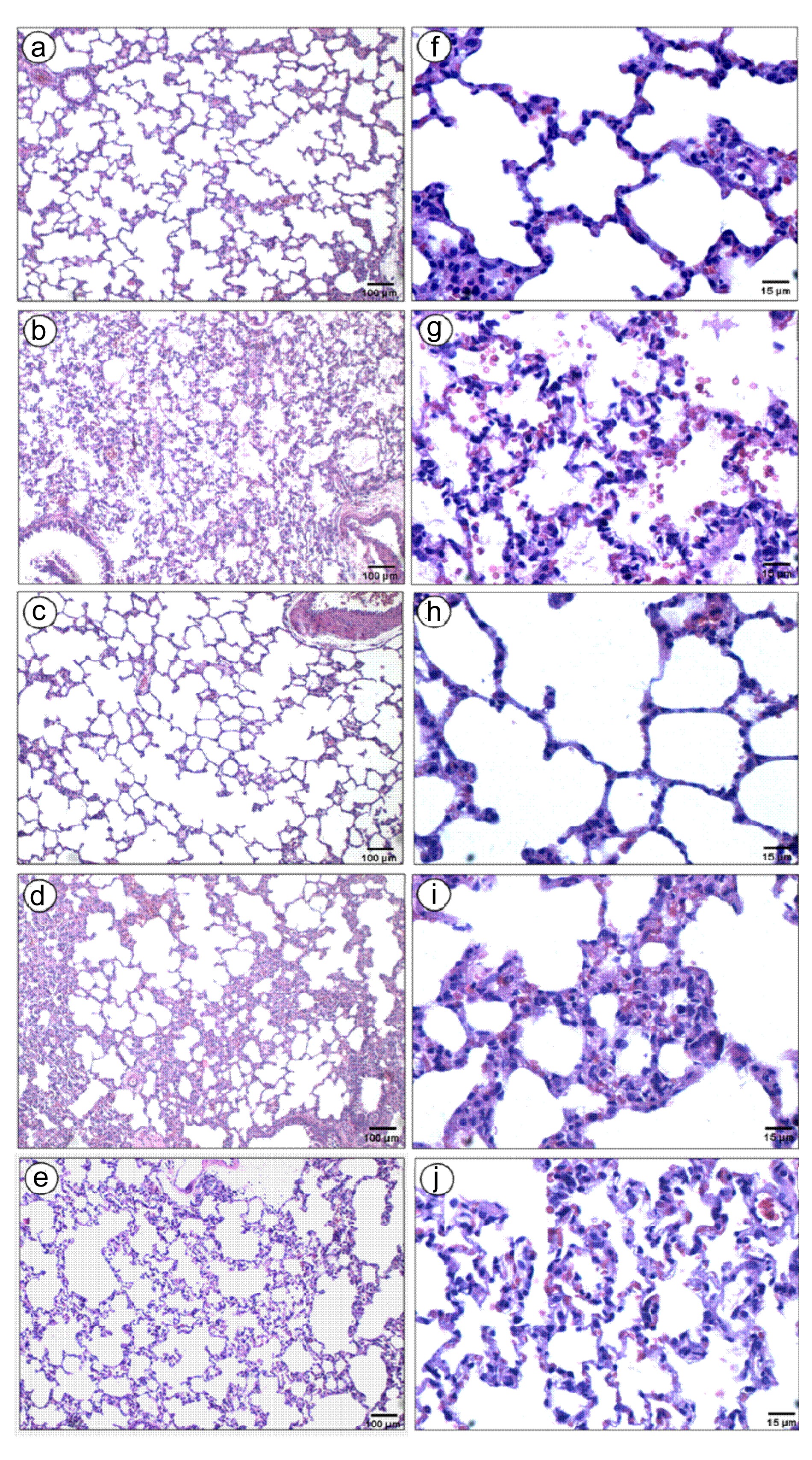
After sacrifice, we observed gross lesions in the lungs of both the male and female rats of the positive control group, and groups treated with PMs. We noticed that the distribution of lesions in the 350 μg PM-treated group was much less severe than that in the positive control group. The lesions consist of the reduction of alveolar ducts and sacs and the thickening of pulmonary interstitial tissues (Figs.
1a, 1d, 1e, 1h, 1i and 1j) compared with the control (Figs.
1a and 1f). In addition, the lung tissue of the positive control group of male rats was seriously damaged (Figs.
1b and 1g) compared to that of the negative control group in the histology by hematoxylin-eosin (H&E) staining, indicating reliability and consistency of our intra-tracheal instillation technique. In male rats, the lung tissues appeared to be normal in the 10 μg PM-treated group (Figs.
1c and 1h); they started to be damaged in the 100 μg (Figs.
1d and 1i) and were worse in the 350 μg PM-treated groups (Figs.
1e and 1j).
Fig.1
Histology of male rat lungs (n=10) stained with hematoxylin-eosin (H&E)
(a, f) Negative control; (b, g) Positive control; (c, h) PM 10 μg; (d, i) PM 100 μg; (e, j) PM 350 μg
In female rats, the lung tissues were injured by 10 μg of PM treatments (Figs.
2b and 2f) compared to the negative control group (Figs.
2a and 2e) and were more severe in 100 μg (Figs.
2c and 2g) and 350 μg (Figs.
2d and 2h) PM-treated groups.
Fig.2
Histology of female rat lungs (n=10) stained with hematoxylin-eosin (H&E)
(a, e) Negative control; (b, f) PM 10 μg; (c, g) PM 100 μg; (d, h) PM 350 μg
4. Discussion
This study measured the level of PMs in working areas of six ceramic factories in Lampang Province, Thailand. Results of the measurement showed that the mean values of 8-h average PM levels from six factories ranged from 33.22 to 193.17 μg/m
3 (Table
1). The level of PMs depended on structure, ventilation, and the different departments of the factories including the season of the collecting period. A lot of departments in the factories exhibited relatively high concentration (>100 μg/m
3; Table
1) of PMs, e.g., shaping, grading, casting, coating, and decorating departments. Factory A had a low level of PMs in all their three departments (55 μg/m
3 or less). However, we were not allowed to collect PMs in other areas of this factory; therefore, we cannot give the reason for this event. Factory D also had a low level of PMs (<55 μg/m
3) in all of their departments because the PMs were collected during the rainy season (September), in which the humidity is the highest of the year. Moreover, there were many air blowers on the roof to help the ventilation although factory D (PMs can spread to every part of the factory) had no partition between various departments. Factory E showed the highest mean level of PMs because PM level was very high in the shaping area (523 μg/m
3). In addition, the PM level in every department of factory E tended to be higher than the other factories. It is observed that this factory had a really closed circulation system, there were no windows and the ventilation inside the factory was not very good at all. Factories B, C, and F had no difference in their PM levels although they were different in their sizes. In this study, the results demonstrated that monthly average 8-h samples of PMs taken from four out of the six ceramic factories were higher than 100 μg/m
3.
Exposure to PMs especially PM2.5 and PM10 can increase the risk of respiratory health problems with both short-term and long-term effects (Baccini et al.,
2011; Cakmak et al.,
2012; Hampel et al.,
2012; Salam et al.,
2012). To prevent workers from inhaling high amounts of PMs, wearing masks are strongly recommended during working hours. In addition, natural air ventilation can help to reduce these PM levels. Therefore, factory owners should be concerned and take responsibility to safeguard their employees.
In the elemental analysis of PM, two groups of elements were identified which belong to the high and low concentrations of chemical elements, respectively. Among the high-concentration group, iron is the most interesting one. Iron plays a crucial role in various metabolic processes. However, there is some evidence for the carcinogenicity of iron that this metal possibly triggers hydroxyl radical formation which leads to DNA damage (Gu et al.,
2011; Vijayavel et al.,
2012). Since the concentration of iron is very high in PM extracts (about 3800 mg/kg), the breathing in of PM-contaminated air needs to be strictly controlled.
Among the low-concentration group, a lot of heavy metals were detected in PM extracts. Although the levels of heavy metals were low in units of milligram per kilogram, these metals are known to play roles in carcinogenesis (Yang,
2011; Martinez-Zamudio and Ha,
2011). Lead was high in this group and it can lead to the cancers of stomach, intestine, renal, lung, and myeloma and leukemia (van Bemmel et al.,
2011; Ilychova and Zaridze,
2012). Cadmium is known to be a mutagen in mammalian, causing DNA damage, and can be associated with cancers of prostate, renal, and lung (Li et al.,
2011; Luo et al.,
2011; Osipov et al.,
2011; Adams et al.,
2012). The other two metals which are known to relate to the development of cancers especially the speed-up of tumor growth are chromium and zinc (Rudolf and Cervinka,
2006; Beyersmann and Hartwig,
2008; Chadha et al.,
2010; Gumulec e
t al.,
2011). In humans, these metals have been associated with a more rapid progression of cancers of breast, colon, rectum, ovary, lung, pancreas, bladder, and leukemia (Franklin and Costello,
2009; Galanis et al.,
2009; Chadha et al.,
2010).
In fact, not all heavy metals are toxic and not all toxic heavy metals have the same toxicity. Therefore, many countries differentiate heavy metals into three classes by ranking their toxicity levels. This study revealed heavy metals of the three classes, i.e., class I: cadmium; class II: arsenic, nickel, and selenium; and, class III: lead, chromium, copper, and manganese. The heavy metal of class I is most toxic and harmful, whereas the ones of class III are least toxic. In the present finding, PM extracts had higher cadmium concentration (17 mg/kg) than the World Health Organization (WHO) annual average guideline (5 ng/kg). Moreover, chromium concentrations in all samples (39 mg/kg) were higher than 2.5 ng/kg of the WHO guideline, which corresponds to an excess of lifetime risk of 1:10 000 (WHO,
2000). The metals found in the PM extracts may come from raw materials used in ceramic factories. Therefore, it is necessary to measure and control the amount of chemical elements in raw materials for the workers, helping to avoid carcinogen exposure.
PMs collected from the different factories contain condensed organic matters that are extractable by organic solvents (Vinitketkumnuen et al.,
2002). A number of studies have shown that the organic extractable matters from air particles and different combustion sources are carcinogenic in animals (Hueper et al.,
1962) and mutagenic in short-term bioassay tests (Alfheim et al.,
1983). However, our results showed that dichloromethane extracts of PMs, collected in ceramic factories, were not mutagenic to
S. typhimurium strain TA 100 with metabolic (S9 mix) activation as compared to the positive control. The metabolic activation (S9) is a biotransformation of relatively inert chemicals to high metabolites with numerous chemically induced toxicities (Patricia,
1994). However, this experiment was done in the bacterial model, and there are more complicated processes in an
in vivo system.
The present findings indicate that PM at a high dose (350 μg) damages the lung of both male and female rats (Figs.
1 and
2). However, PM at a lower amount (10 μg) does not affect the lung in the histology of a male rat, whereas PM at dosages of 100 μg starts to affect it. In contrast, the lungs of the female rats started to be damaged with PM at dosages of 10 μg and its damage was more severe at 350 μg PMs (Fig.
2). Therefore, this lung response was attenuated in male rats. These findings indicate that sexes may be correlated with the level of lung damage by intra-tracheal instillation of PMs. It has been reported that intranasal instillation of fine PM (2.5 μm) can induce acute lung inflammation in mice (Riva et al.,
2011). In addition, Budinger et al. (
2011) revealed severe lung damage in mice which received ambient PMs via intra-tracheal instillation. Obviously, the present study also demonstrated in rats that ceramic PMs induced histological changes of lung tissues, which consist of the reduction of alveolar duct and sac spaces, and increased interstitial connective tissues (alveolar septa) as compared to the normal control lung tissues (Figs.
1 and
2). Taken all together, the present study clarified that PMs can primarily induce the histological changes of rodent lungs. Since the expressions of some cancer markers have been reported in the lung tissue receiving PMs (Laskin et al.,
2010; Budinger et al.,
2011; Riva et al.,
2011), it is necessary to examine the effects of ceramic PM on the expression of lung cancer markers as the next step.
In conclusion, although PM extracts consist of high amounts of iron and a detectable amount of lead, cadmium, chromium, and zinc, they are not mutagenic for
S. typhimurium strain TA 100. In rats, the induction effect of ceramic PMs on histological changes of the lung tissues is more sensitive in females than in males. These structural changes may imply the possibility of PM-induced lung cancer whose mechanism remains to be elucidated. Importantly, ceramic industries must seriously take note of the necessity for different ways to protect employees from the risk of high PM inhalation.
Acknowledgements
We would like to thank Dr. Thiravud KUHAPREMA, Director of National Cancer Center, Thialand, for his efforts in research funding, and the Lampang Provincial Labor Protection and Welfare Office for their cooperations in the PM collection process. We would also like to thank Prof. Hisatake KONDO, overseas visiting professor of the Anatomy Department, Faculty of Medicine, Khon Kaen University, Thialand, for proofreading through this article.
* Project supported by the Oncological Society of Thailand under the Royal Patronage of Her Majesty the Queen (No. RE53006)Compliance with ethics guidelines Duriya FONGMOON, Surathat PONGNIKORN, Aphiruk CHAISENA, and Sitthichai IAMSAARD declare that they have no conflict of interest.References
[1] Adams, S.V., Passarelli, M.N., Newcomb, P.A., 2012. Cadmium exposure and cancer mortality in the Thrid National Health and Nutrition Examination Survey cohort.
Occup Environ Med, 69(2):153-156.


[2] Alfheim, I., Lfroth, G., Mller, M., 1983. Bioassay of extracts of ambient particulate matter.
Environ Health Perspect, 47:227-238.
[3] Baccini, M., Biggeri, A., Grillo, P., 2011. Health impact assessment of fine particle pollution at the regional level.
Am J Epidemiol, 174(12):1396-1405.
[4] Beyersmann, D., Hartwig, A., 2008. Carcinogenic metal compounds: recent insight into molecular and cellular mechanisms.
Arch Toxicol, 82(8):493-512.
[5] Budinger, G.R., McKell, J.L., Urich, D., 2011. Particulate matter-induced lung inflammation increases systemic levels of PAI-1 and activates coagulation through distinct mechanisms.
PLoS ONE, 6(4):e18525
[6] Cakmak, S., Dales, R.E., Coates, F., 2012. Does air pollution increase the effect of aeroallergens on hospitalization for asthma?.
J Allergy Clin Immunol, 129(1):228-231.
[7] Chadha, V.D., Gerg, M.L., Dhawan, D., 2010. Influence of extraneous supplementation of zinc on trace elemental profile leading to prevention of dimethylhydrazine-induced colon carcinogenesis.
Toxicol Mech Meth, 20(8):493-497.
[8] Franklin, R.B., Costello, L.C., 2009. The important role of the apoptotic effects of zinc in the development of cancers.
J Cell Biochem, 106(5):750-757.
[9] Galanis, A., Karapetsas, A., Sandaltzopoulos, R., 2009. Metal-induced carcinogenesis, oxidative stress and hypoxia signaling.
Mutat Res/Genet Toxicol Environ Mutagen, 674(1-2):31-35.
[10] Gu, Y., Hua, Y., He, Y., 2011. Iron accumulation and DNA damage in a pig model of intracerebral hemorrhage.
Acta Neurochir Suppl, 111:123-128.
[11] Gumulec, J., Masark, M., Krzkov, S., 2011. Molecular mechanisms of zinc in prostate cancer.
Klin Onkol, 24(4):249-255.
[12] Hampel, R., Breitner, S., Schneider, A., 2012. Acute air pollution effects on heart rate variability are modified by SNPs involved in cardiac rhythm in individuals with diabetes or impaired glucose tolerance.
Environ Res, 112:177-185.
[13] Hueper, W.C., Kolin, P., Tabor, E.C., 1962. Carcinogenic bioassays on air pollutants.
Arch Pathol, 74:89-116.
[14] Ilychova, S.A., Zaridze, D.G., 2012. Cancer mortality among female and male workers occupationally exposed to inorganic lead in the printing industry.
Occup Environ Med, 69(2):87-92.
[15] Laskin, D.L., Mainelis, G., Turpin, B.J., 2010. Pulmonary effects of inhaled diesel exhaust in young and old mice: a pilot project.
Res Rep Health Eff Inst, 151:3-31.
[16] Lauer, F.T., Mitchell, L.A., Bedrick, E., 2009. Temporal-spatial analysis of U.S.-Mexico border environmental fine and coarse PM air sample extract activity in human bronchial epithelial cells.
Toxicol Appl Pharmacol, 238(1):1-10.
[17] Li, R., Yuan, C., Dong, C., 2011. In vivo antioxidative effect of isoquercitrin on cadmium-induced oxidative damage to mouse liver and kidney.
Naunyn-Schmiedebergs Arch Pharmacol, 383(5):437-445.
[18] Luo, J., Hendryx, M., Ducatman, A., 2011. Association between six environmental chemicals and lung cancer incidence in the United States.
J Environ Public Health, 2011:463701
[19] Mantecca, P., Farina, F., Moschini, E., 2010. Comparative acute lung inflammation induced by atmospheric PM and size-fractionated tire particles.
Toxicol Lett, 198(2):244-254.
[20] Maron, D.M., Ames, B.N., 1983. Revised methods for the
Salmonella mutagenicity test.
Mutat Res/Environ Mutagen Relat Subj, 113(3-4):173-215.
[21] Martinez-Zamudio, R., Ha, H.C., 2011. Environmental epigenetics in metal exposure.
Epigenetics, 6(7):820-827.
[22] Nishi, K., Morimoto, Y., Ogami, A., 2009. Expression of cytokine-induced neutrophil chemoattractant in rat lungs by intratracheal instillation of nickel oxide nanoparticles.
Inhal Toxicol, 21(12):1030-1039.
[23] Osipov, A.N., Riabchenko, N.I., Ivannik, B.P., 2011. DNA damage in thymocytes of mice under combined acute whole body exposure to cadmium ions and gamma-radiation.
Radiats Biol Radioecol, 51(3):315-320.
[24] Patricia, E.L., 1994. Reactive metabolites and toxicity. Introduction to Biochemical Toxicology. Appleton & Lange,Norwalk, Conn. :219
[25] Raunroadroong, N., Daoprasert, K., Srisukho, S., 2009.
Cancer Incidence in Northern Thailand 20032007, Lampang Cancer Center,:10-29.
[26] Riva, D.R., Magalhaes, C.B., Lopes, A.A., 2011. Low dose of fine particulate matter (PM2.5) can induce acute oxidative stress inflammation and pulmonary impairment in healthy mice.
Inhal Toxicol, 23(5):257-267.
[27] Rudolf, E., Cervinka, M., 2006. The role of intracellular zinc in chromium(VI)-induced oxidative stress, DNA damage and apoptosis.
Chem Biol Interact, 162(3):212-227.
[28] Salam, M.T., Byun, H.M., Lurmann, F., 2012. Genetic and epigenetic variations in inducible nitric oxide synthase promoter particulate pollution and exhaled nitric oxide levels in children.
J Allergy Clin Immunol, 129(1):232-239.
[29] Tokiwa, H., Horikawa, K., Sera, N., 1992. Influence for the microsomal inducer and the incubation system on mutagenicity of complex mixtures.
Mutat Res/Rev Genet Toxicol, 276(1-2):139-144.
[30] Toya, T., Fukuda, K., Takaya, M., 2001. Lung lesions induced by intratracheal instillation of vanadium pentoxide powder in rats.
Ind Health, 39(1):8-15.
[31] van Bemmel, D.M., Boffetta, P., Liao, L.M., 2011. Comprehensive analysis of 5-aminolevulinic acid dehydrogenase (ALAD) variants and renal cell carcinoma risk among individuals exposed to lead.
PLoS ONE, 6(7):e20432
[32] Vijayavel, K., Downs, C.A., Ostrander, G.K., 2012. Oxidative DNA damage induced by iron chloride in the larvae of the lace coral Pocillopora damicornis.
Comp Biochem Physiol C Toxicol Pharmacol, 155(2):275-280.
[33] Vinitketkumnuen, U., Kalayanamitra, K., Chewonarin, T., 2002. Particulate matter PM 10 & PM 2.5 levels and airborne mutagenicity in Chiang Mai Thailand.
Mutat Res/Genet Toxicol Environ Mutagen, 519(1-2):121-131.
[34] World Health Organization (WHO), 2000. Air Quality Guidelines for Europe, 2nd Edition.
WHO Regional Publications, European Serials, No. 91, :
[35] Yang, M., 2011. A current global view of environmental and occupational cancers.
J Environ Sci Health C Environ Carcinog Ecotoxicol Rev, 29(3):223-249.


Open peer comments: Debate/Discuss/Question/Opinion
<1>